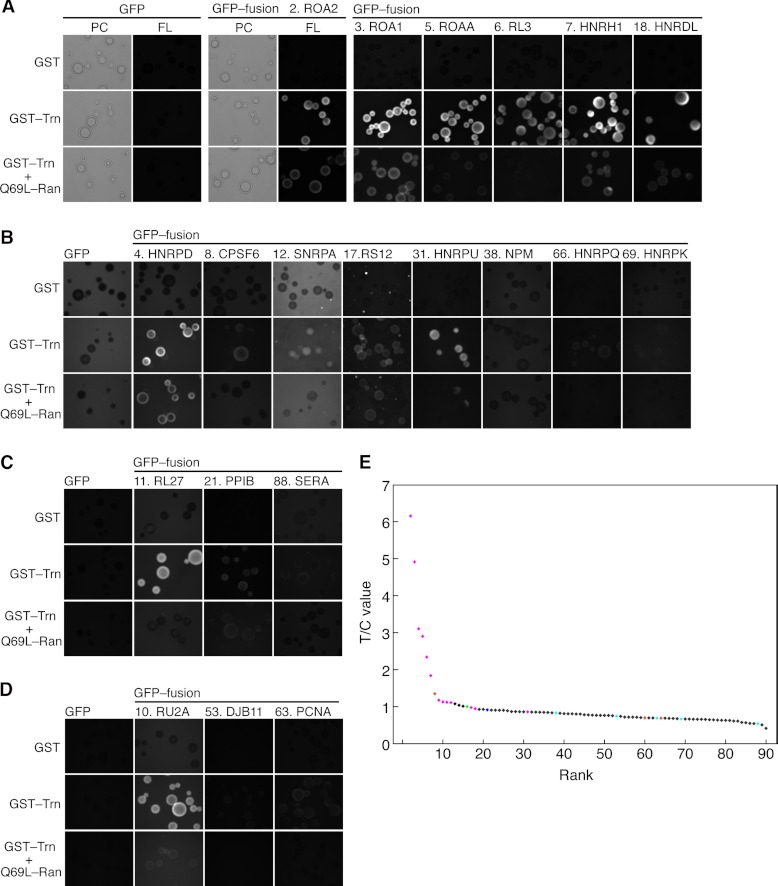
Fig. 2.
Interaction of candidate cargoes with Trn. A–D, interactions examined via bead halo assay. Bacterial extracts containing GFP-fusion proteins were mixed with GSH-Sepharose beads coated with GST or GST-Trn fusion proteins and then observed via fluorescence microscopy. Q69L-Ran, which inhibits the Trn-cargo specific interaction, was added as indicated. The protein numbering is identical to that in Table I. The images within each panel are comparable. PC, phase contrast; FL, fluorescence micrograph. A, B, the assay and microscopy conditions were appropriate for proteins expressed at high levels in E. coli. B, images taken with the same exposure time as in A were enhanced. C, D, proteins expressed at lower levels. The GFP moiety concentrations were half (C) and one-fifth (D) of those in A and B. Accordingly, the exposure time was longer. E, T/C values predict Trn cargoes. Proteins quantified via MS were ranked in descending T/C value order (Table I). Horizontal, protein rank; vertical, T/C value. Magenta, protein (or segment, Figs. 3C and 3D) that bound to Trn in the bead halo assays; blue, unclear; cyan, not bound; orange, not examined here but reported to be Trn cargo; green, IMB1; black, others. RA1L3 (sequence from pseudogene) is not shown.

