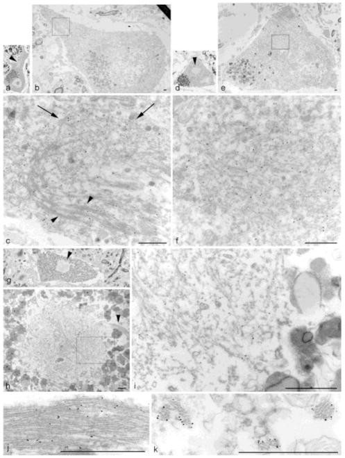
Figure 5. TDP-43 nanogold immuno-EM on lumbar spinal cord sections from 3 ALS cases.
Case #1 (a–f) showing an elongated inclusion with bundles of randomly oriented filaments (a–c) and a compact inclusion with granulo-filamentous material (d–f). (a) Semi-thin section of a large motor neuron shows a low power view of the inclusion (arrowhead) (60X). (b) Same cell re-embedded for EM (1300X). (c) Higher magnification of (b). Note bundles of filaments at bottom (arrowheads) and randomly oriented filaments toward top (arrows) (14,600X). (d) Semi-thin section of a large motor neuron with a round inclusion (arrowhead) (60X). (e) Same cell re-embedded for EM (1300X). (f) Higher magnification of boxed area in (e) (16,500X). Note the specificity of the TDP-43-linked nanoparticles by the heaviliy decorated fibrils in the central portion of the panel compared to the tissue in the lower left with minimal particles. Case #2 (g–j) showing a compact inclusion associated with a bundle of filaments (g–i) and bundle of filaments from another section (j). (g) Semi-thin section of a large motor neuron with a compact inclusion (arrowhead) (60X). (h) Same cell re-embedded for EM, showed a tight bundle of filaments (arrowhead) coming out of a round aggregate of granulo-filamentous material (4000X). (i) Higher magnification of boxed area in (h) (22,600X). (j) Higher magnification of a TDP-43 nanogold decorated bundle of filaments from another section (43,400X). Case #3 (k) Cross-section of TDP-43 nanogold decorated filament bundles (5,1400X). (scale bars = 1μm)

