Fig 4.

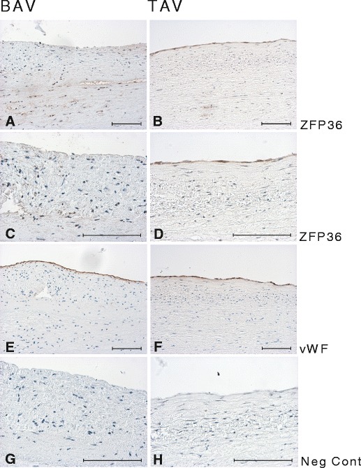
Immunostaining of ZFP36 in non-dilated aorta of BAV (a, c) and TAV (b, d) patients. e, f vWF staining in BAV and TAV, respectively; g, h negative control. Scale bar = 100 μm

Immunostaining of ZFP36 in non-dilated aorta of BAV (a, c) and TAV (b, d) patients. e, f vWF staining in BAV and TAV, respectively; g, h negative control. Scale bar = 100 μm