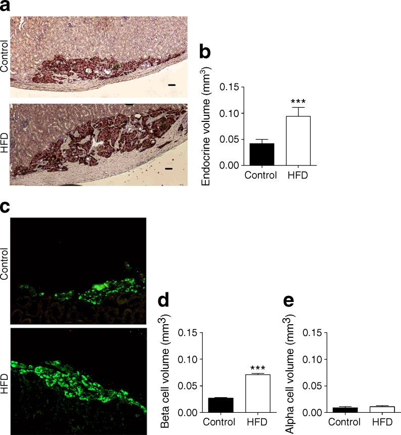
Fig. 2.
(a) Representative sections of paraffin-embedded human grafts stained with the endocrine marker chromogranin A (DAB, brown) in HFD and control-diet mice (scale bar, 50 μm). (b) Morphometric analysis for total endocrine volume (chromogranin A) (HFD [n = 6] vs control [n = 7], >196 sections/group; linear mixed model, see Methods, ***p < 0.001). (c) Sections of paraffin-embedded human grafts stained with the beta cell marker C-peptide (green). (d) Morphometric analysis for beta cell volume (HFD [n = 6] vs control [n = 7], ***p < 0.001). (e) Morphometric analysis for alpha cell volume (HFD [n = 6] vs control [n = 7])

