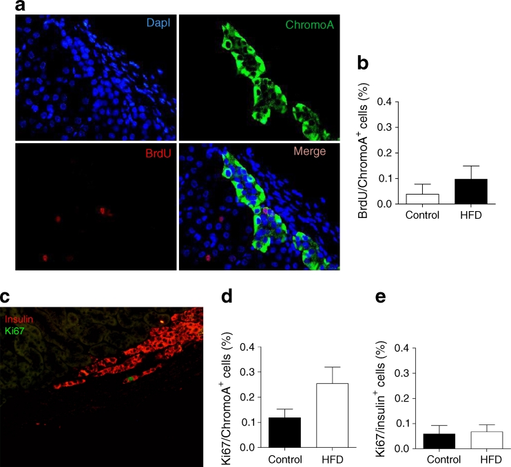
Fig. 4.
(a) Proliferation was determined on sections of human grafts at 12 weeks following an overnight incubation with BrdU (nuclei in blue, BrdU in red, chromogranin A [chromoA] in green or merge). (b) BrdU/chromogranin A endocrine-positive cell quantification in human islet grafts (HFD [n = 4] vs control [n = 5]). (c) Proliferation with Ki67 in beta cells (image shows merge of insulin in red, Ki67 in green). Quantification of (d) Ki67/chromogranin A and (e) Ki67/insulin-positive cells in human islets grafts (HFD [n = 4] vs control [n = 5]; linear mixed model, see Methods, p = 0.08)

