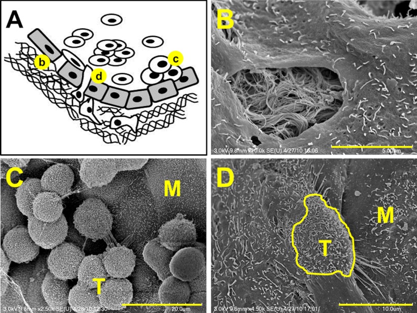
FIGURE 1.
Model of epithelial ovarian cancer metastasis. A, cells shed from the primary tumor circulate in ascites fluid, interact with peritoneal mesothelial cells, induce mesothelial cell retraction, and adhere avidly to the submesothelial three-dimensional collagen matrix wherein they anchor and proliferate to form secondary lesions. Scanning electron micrographs (SE) depicting events in metastasis (b, c, and d) are shown in the following panels. B, scanning electron micrographs of retracted mesothelial cells showing the underlying three-dimensional collagen matrix. Scale bar, 5 μm. C, scanning electron micrographs of EOC tumor cells (T) adherent to peritoneal tissue in a tissue explant. M designates mesothelial cells. Scale bar, 20 μm. D, scanning electron micrographs of EOC tumor cell (T; outlined in yellow) intercalated between peritoneal mesothelial cells (M) in a tissue explant. Scale bar, 10 μm.

