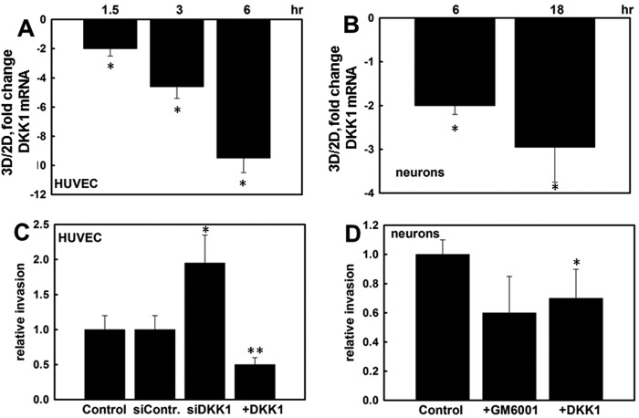
FIGURE 7.
Three-dimensional CI regulates DKK1 expression and invasion in endothelial cells and primary cortical neurons. Human umbilical vein endothelial cells (HUVEC) (A) and E17 primary cortical neurons (B) were cultured on three-dimensional (3D) CI or planar (two-dimensional (2D)) collagen I for the indicated periods of time. RNA was extracted, cDNA was synthesized, and real time RT-PCR was performed to detect DKK1 RNA expression. Ratios of DKK1 RNA expression were found with the 2−ΔΔCt method. The averaged ratio of DKK1 expression in three-dimensional CI versus two-dimensional CI from three independent experiments is depicted in the histograms ±S.D. (error bars). * designates p < 0.0005. C, endothelial cells were transiently transfected with control siRNA (siContr), DKK1-specific siRNA (siDKK1), or a DKK1-expressing plasmid (+DKK1) as indicated followed by evaluation of the ability to invade three-dimensional CI gels. Invasion of untreated control cells was arbitrarily set as 1, and other values were calculated accordingly. The average of four independent experiments ±S.D. (error bars) is presented. * designates p < 0.05, and ** designates p < 0.005 relative to control. D, the ability of E17 cortical neurons to invade three-dimensional CI gels was assessed in the presence and absence of 10 μm GM6001 or 0.33 mg/ml exogenous DKK1 as indicated. The average of three independent experiments ±S.D. (error bars) is presented. * designates p < 0.05 relative to untreated controls. Note that comparison of invasion in the presence of GM6001 resulted in p = 0.06.

