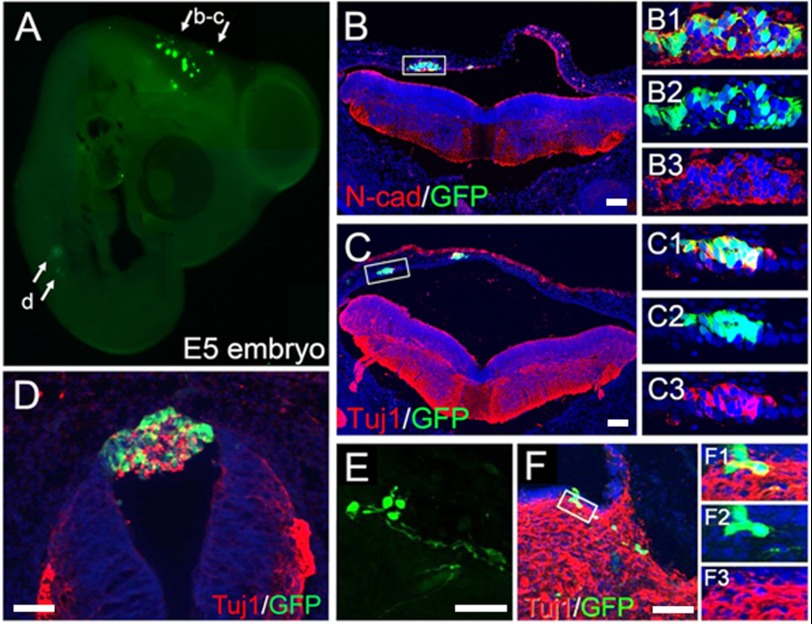
FIGURE 2.
A, transplanted human pNSCs at E5. Many pNSCs formed clusters in the dorsal wall of the mesencephalon and expressed N-cadherin (N-cad; B) and Tuj1 (C). Enlarged images of the boxed areas in B and C are shown in B1–B3 and C1–C3, respectively. D, pNSCs in the dorsal spinal cord also formed cell clusters with Tuj1 expression. E and F, transplanted cells integrated into the ventral mesencephalon. Cells exhibited long neurites (E) and expressed Tuj1 (F). Enlarged images of the boxed area in F are shown in F1–F3. Scale bars = 100 μm.

