FIGURE 3.
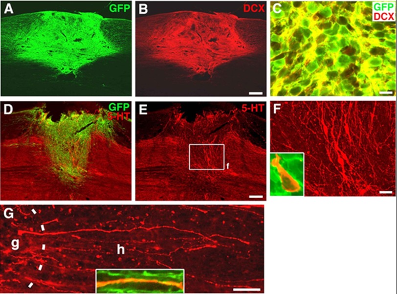
Survival and neuronal differentiation of human pNSCs after transplantation to the lesion site of an adult rat spinal cord. Immunohistochemical analysis revealed excellent survival of transplanted pNSCs expressing GFP (A) that co-localized with the early neuronal marker doublecortin (DCX) (B and C). GFP and 5-HT immunolabeling demonstrated 5-HT-positive neurons and their processes within the graft site (D–F). F is a higher magnification of the boxed area in E. The inset shows co-localization of a 5-HT neuron with GFP expression, confirming derivation from graft cells. Many linear 5-HT fibers extended into the host spinal cord from the graft site (G). The dashed line indicates the graft (g)-host (h) interface. The inset shows co-localization of a 5-HT axon with GFP. Scale bars = 230 μm (A and B), 10 μm (C), 310 μm (D and E), 30 μm (F), and 110 μm (G).

