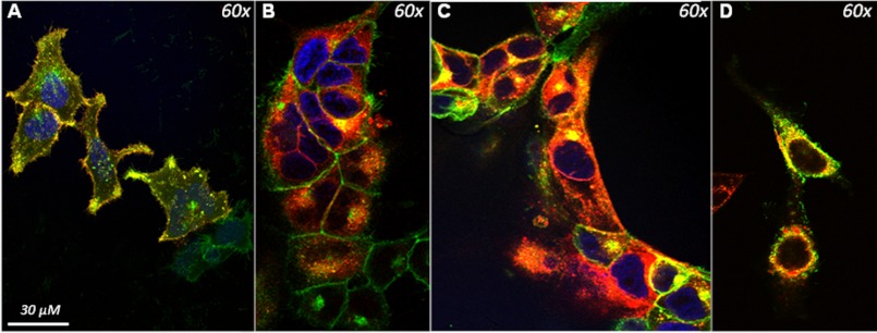
FIGURE 1.
Co-localization of the GHS-R1a receptor with candidate GPCRs in Hek293A cells. Hek293a cells stably expressing the GHS-R1a receptor as an EGFP fusion protein were transduced with lentiviral vectors expressing candidate GPCRs as RFP fusion proteins. Co-localization of fluorescence was analyzed using confocal microscope and is indicated by yellow color overlap. A, co-localization (yellow) of the D1 receptor (red) with the GHS-R1a receptor (green) was observed. B, co-localization of the MC3 receptor (red) with the GHS-R1a (green) receptor was only observed intracellularly. C, co-localization of the 5-HT2C receptor (red) with the GHS-R1a receptor (green) was demonstrated both intracellularly and on the membrane. D, the edited 5-HT2C isoform, 5-HT2C-VSV receptor (red), also demonstrated co-localization with the GHS-R1a receptor mainly in the intracellular space.

