FIGURE 3.
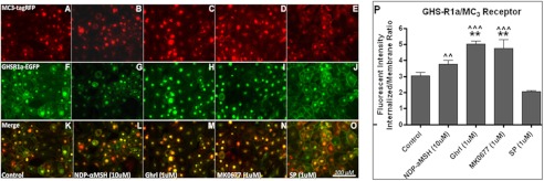
Co-internalization of the GHS-R1 receptor with the MC3 receptor. Hek293A cells stably expressing GHS-R1a-EGFP were transduced with viral vectors expressing lvMC3-RFP. A–O, expression of the MC3 receptor (red), the GHS-R1a receptor (green), and co-localization (yellow) could be observed 48 h after transduction (A, F, and K). The cells were incubated for 60 min with 10 μm of the specific MC3-agonist, [Nle4,d-Phe7]-α-MSH (B, G, and L), with 1 μm ghrelin (C, H, and M), with 1 μm MK0677 (D, I, and N), or with 1 μm of [-Arg1,d-Phe5,d-Trp7,9,Leu11]-substance P (SP; E, J, and O). Images were made using a CKX41 inverted microscope (Olympus) with a XM10 camera and Cell̂Fv3.3 software. P, quantified agonist-mediated co-internalization is depicted. Statistical significance was analyzed using ANOVA followed by Bonferroni multiple comparison test; statistical significance of agonist-mediated co-internalization compared with control is notated as follows: ***, p < 0.001; **, p < 0.01; and *, p < 0.05. Statistically significant difference compared with [d-Arg1,d-Phe5,d-Trp7,9,Leu11]-substance P is notated as follows: ^^^, p < 0.001; ^^, p < 0.01; and ^, p < 0.05.

