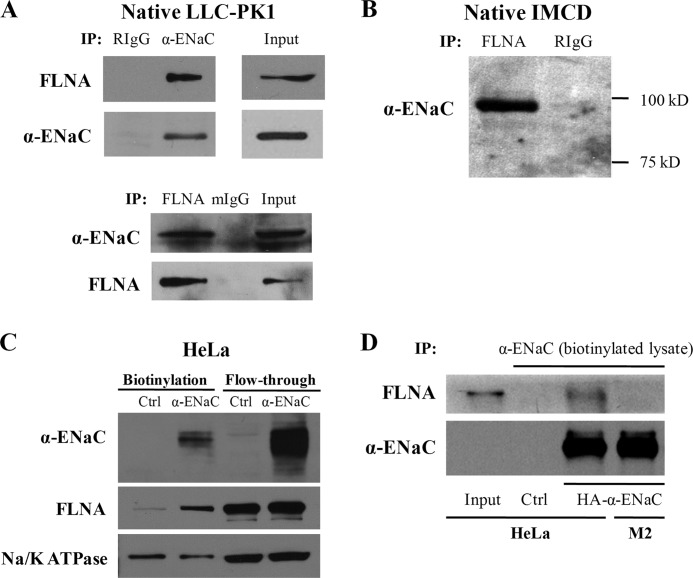
FIGURE 3.
Interaction between endogenous ENaC and filamin A by co-IP. Data shown here are representative of those from three to four independent experiments. A, LLC-PK1 cell lysates were used for reciprocal co-IP using anti-α-ENaC (Calbiochem) and anti-FLNA E-3 for IP and IB as indicated. Nonimmune serum rabbit IgG (RIgG) and mouse IgG (mIgG) were used as controls. B, cell lysates from IMCD cells were precipitated with anti-FLNA H-300 and RIgG as control. Immunoprecipitated proteins were analyzed by WB using anti-α-ENaC antibody (Calbiochem). C, biotinylation was performed on HeLa cells transfected with either human α-ENaC or empty pcDNA3.1 plasmid (Ctrl). Biotinylated proteins were subjected to SDS-PAGE and detected by antibody against ENaC, FLNA, or Na/K-ATPase (as a control). D, biotinylation was performed on HeLa and M2 cells transfected with human α-ENaC or empty pcDNA3.1 plasmid (HeLa only, Ctrl). Biotinylated proteins were collected using the monomeric avidin kit (Pierce) and proceeded to co-IP assays with α-ENaC antibody. Precipitated proteins were immunoblotted with FLNA H-300 or α-ENaC antibody.

