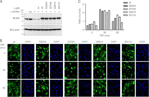
FIGURE 1.
Generation and characterization of wild type and phosphorylation-deficient p65-expressing bEND.3 cells. A, Western blot analysis of p65 expression in bEND.3 cells transduced with luciferase (luc) control and p65-targeting shRNAs (+). c-Myc-tagged human p65 wild type (wt) and amino acids 205, 276, 281, 311 serine to alanine (SA) mutants were stably introduced into successful p65 knockdown cells. The fusion of p65 with N-terminal c-Myc leads to a shift in molecular weight of 10 amino acids reflected by slower migration of human c-Myc-p65 in SDS-PAGE. IB, immunoblot. B, subcellular localization of p65 variants in bEND.3 in the absence or presence of 10 ng/ml TNF for 30 and 60 min. DAPI staining is included as nuclear reference. Bar, 20 μm. C, semi-automated quantification of cytosolic and nuclear p65 immunofluorescence (n = 128–248 cells, derived from three experiments). Nuclear/cytosolic values of >1 indicate predominantly nuclear p65.

