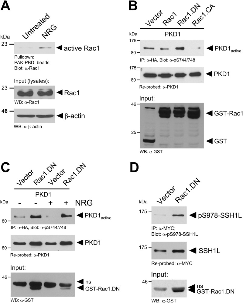
FIGURE 7.
NRG negatively regulates PKD1 via Rac1. A, MCF-7 cells were serum-starved for 24 h and then stimulated with NRG (100 ng/ml). Cells were lysed, and active Rac1 was pulled down. Samples were subjected to SDS-PAGE, transferred to nitrocellulose, and analyzed for Rac1 by immunoblotting with anti-Rac1 (total Rac1) antibody. Cell lysates were also stained with anti-Rac1 antibody to determine total Rac1 (input control). WB, Western blot. B, cells were transfected with HA-tagged PKD1 and GST-tagged Rac1, dominant-negative Rac1 (Rac1.DN), or constitutively active Rac1 (Rac1.CA) as indicated. 24 h after transfection, cells were lysed, and PKD1 was immunoprecipitated (IP; anti-HA antibody). Samples were subjected to SDS-PAGE, transferred to nitrocellulose, and analyzed for PKD1 activity by immunoblotting with anti-phospho-Ser-744/748 PKD antibody (which recognizes PKD1 phosphorylated at Ser-738 and Ser-742). Immunoblots were restained for PKD1 (anti-PKD1 antibody). Cell lysates were stained with anti-GST antibody for overexpression of GST-Rac1. C, MCF-7 cells were transfected with vector control, PKD1, or dominant-negative Rac1 as indicated. After transfection, cells were serum-starved for 16 h and then stimulated with NRG (100 ng/ml) as indicated. Cells were lysed, and PKD1 was immunoprecipitated (anti-HA antibody). Samples were subjected to SDS-PAGE, transferred to nitrocellulose, and analyzed for PKD1 activity by immunoblotting with phospho-Ser-744/748 PKD antibody. For controls, immunoblots were restained for PKD1 (anti-PKD1 antibody). Cell lysates were stained with anti-GST antibody for overexpression of GST-Rac1. D, MCF-7 cells were transfected with Myc-tagged SSH1L and GST-tagged dominant-negative Rac1 as indicated. 24 h after transfection, cells were lysed, and SSH1L was immunoprecipitated (anti-Myc antibody). Samples were subjected to SDS-PAGE, transferred to nitrocellulose, and analyzed for phosphorylation of SSH1L at Ser-978 using anti-Ser-978 SSH1L antibody. For controls, immunoblots were restained for ectopically expressed SSH1L (anti-Myc antibody). Cell lysates were stained with anti-GST antibody for overexpression of GST-Rac1.

