Figure 2.
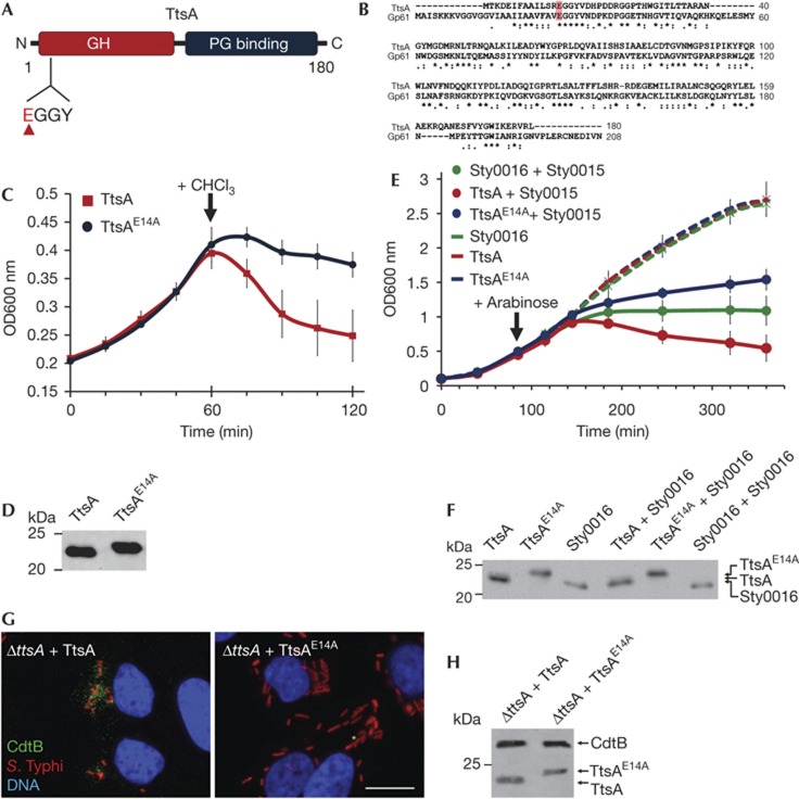
The TtsA enzymatic activity is required for toxin secretion. (A) TtsA domain organization. Indicated are the locations of the GH and PG-binding domains, as well as the catalytic site (EGGY). (B) Amino-acid sequence alignment of TtsA and coliphage N4 Gp61 muramidase. The conserved catalytic glutamate is highlighted. (C) Bacteriolytic activity of TtsA. FLAG-epitope tagged TtsA or TtsAE14A were overexpressed in S. Typhi ΔttsA mutant and at the indicated time 0.3% chloroform (CHCl3). Bacterial lysis was monitored by measuring the OD600 nm of the bacterial cultures. The graph shows the average and s.d.’s of six independent assays. (D) Western blot analysis of the expression levels of the indicated proteins in the strains used in panel (C) in aliquots obtained immediately before adding CHCl3. (E) Holin-assisted bacteriolysis by TtsA. FLAG-tagged TtsA, TtsAE14A or Sty0016 were expressed in a ΔttsA S. Typhi mutant from an arabinose-inducible promoter either by themselves, or along with the Sty0015 holin. At the indicated time, arabinose was added to induce holin/endolysin expression and bacterial lysis monitored by measuring the OD600 nm of the different cultures. The graph shows the average and s.d.’s of six independent assays. (F) Western blot analysis of the expression levels of the different endolysins in the strains used in panel (E) 20 min following the induction with arabinose. (G) The TtsA catalytic activity is required for toxin secretion. Henle-407 cells were infected with a ΔttsA S. Typhi mutant expressing chromosomally encoded FLAG-tagged cdtB complemented with either wild-type TtsA or the catalytic mutant TtsAE14A. Twenty-two hours post infection, infected cells were fixed and stained with a mouse monoclonal antibody directed to the FLAG epitope (to visualize CdtB), a rabbit antibody directed to S. Typhi LPS and DAPI for DNA detection. The bar represents 10 μm. (H) Protein levels in CFU standardized lysates of strains used in panel (G) 22 h after infection of Henle-407 cells. CFU, colony forming units; DAPI, 4,6-diamidino-2-phenylindole; GH, glycosyl hydrolase; LPS, lipopolysaccharide; PG, peptidoglycan; s.d., standard deviation.

