Figure 8.

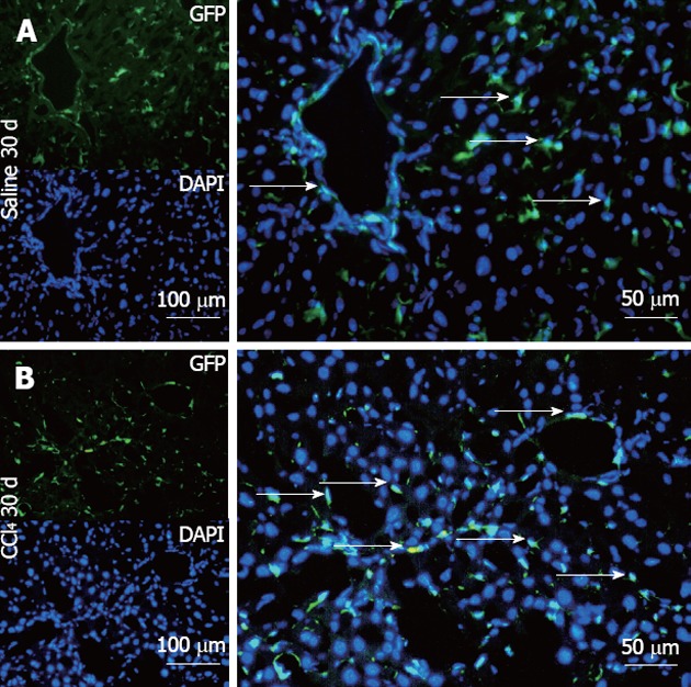
Representative direct fluorescence images of hepatic sections of chimeric animals after transplantation. Liver samples of chimeric animals after transplantation of green fluorescent protein (GFP)+ bone marrow- mononuclear cell, chimeric animals that received saline (Saline 30 d) and chimeric animals that received CCl4 (CCl4 30 d). In panel A (Salina 30 d), the GFP channel shows the presence of green fluorescence; in the 4, 6-diamino-2-phenylindole panel, nuclei are stained blue in the same field above; the merge panel superposes the two images, where GFP+ cells (arrows) can be seen distributed in major hepatic parenchyma and with fusiform morphology. In panel B (CCl4 30 d), GFP+ cells can be seen with greater distribution in the hepatic parenchyma, some fusiform morphology (white arrows) and others with rounded morphology. DAPI: 4,6-diamino-2-phenyl indole.
