Figure 8.
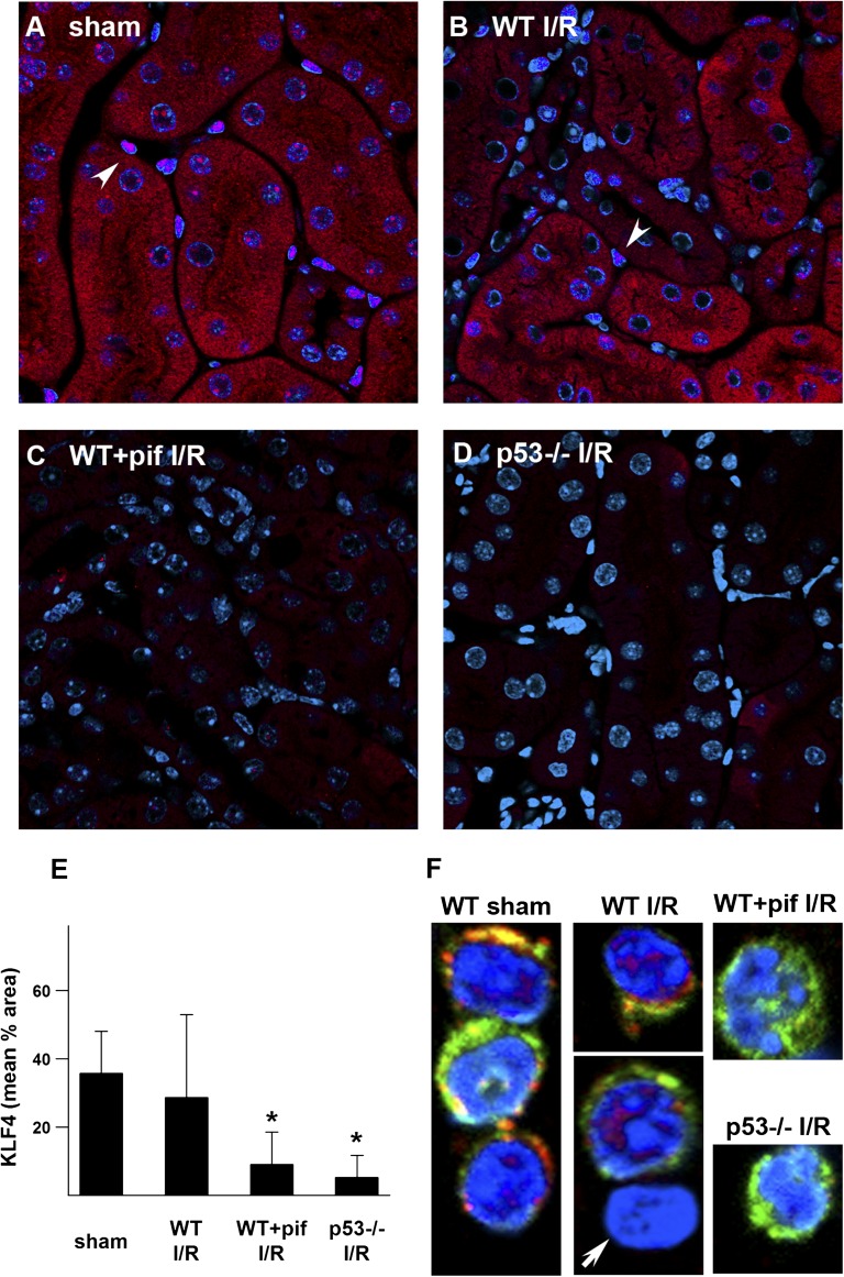
KLF4 expression after kidney IRI. Representative images of KLF4 staining (red; DAPI, blue) are shown for WT mice treated with vehicle control (A and B) or pifithrin-α (C), as well as p53−/− mice (D) under indicated conditions. Arrowheads indicate nuclear KLF4 stain in interstitial cells. (E) Quantitation of tissue KLF4 staining. *P<0.05 versus sham or WT IRI. (F) F4/80+CD11b+ macrophages enriched from kidneys of mice under sham condition or 1 week after IRI are stained for KLF4 (red), CD11b (green), and DAPI (blue). Arrow indicates CD11b negative cell (nonmacrophage) that also lacks KLF4 staining. DAPI, 4',6-diamidino-2-phenylindole; I/R, ischemia-reperfusion injury; pif, pifithrin-α.

