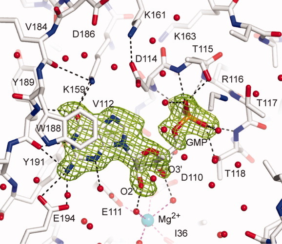
Figure 2.

View of the active site. The 2Fo-Fc electron density maps are contoured at 1.5 σ around the GMP in the chain B of WT2 crystal form of Sc-HGPRT. The hydrogen bond network around the GMP molecule is drawn. The hexa-coordinated magnesium is drawn as a sphere colored in cyan. [Color figure can be viewed in the online issue, which is available at wileyonlinelibrary.com.]
