
Figure 3. Similarity in Regions of Connectivity Loss in Early AD and Cognitively Normal Elderly with Increased Brain Amyloid Binding.
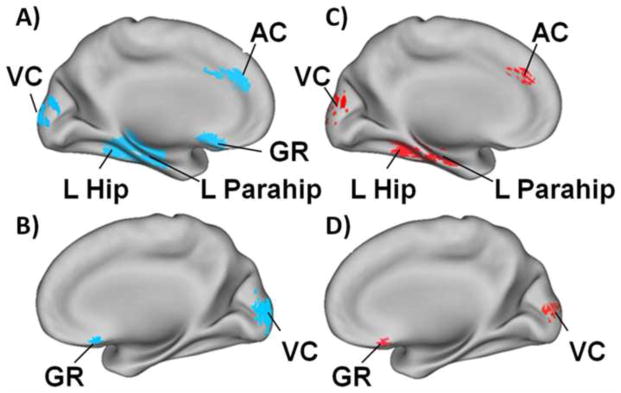
Figure 3A and 3B: Resting state functional connectivity is significantly decreased in early Alzheimers disease. Using the precuneus as the seed region there is less functional connectivity with the left hippocampus (L Hip), left parahippocampus (L Parahip), anterior cingulate cortex (AC) and gyrus rectus (GR) and increased connectivity with visual cortex (VC). Figure 3C and 3D: Again using the precuneus as the seed region, the same pattern of rs-fMRI abnormalities was found in cognitively normal persons with elevated amyloid binding on PIB-PET. The regions with decreased functional connectivity are shown in blue and those with increased connectivity are shown in red. Adapted from Sheline et al (68).