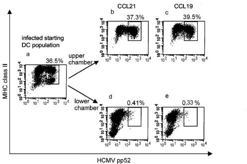
FIG. 1.
HCMV Ag-positive DCs do not migrate toward lymphoid chemokines. DCs, infected with HCMV TB40/E at an MOI of 10 for 24 h and then matured with LPS/IFN-γ/TNF-α for a further 24 h, were placed into transwell migration chambers which contained a 200-ng/ml concentration of either CCL21 (b and d) or CCL19 (c and e) in the lower chambers. The experiment was run for 4 h, and samples of the input DCs (a), DCs which did not migrate (b and c), and those which migrated into the lower chambers (d and e) were analyzed for the rate of HCMV infection by immunofluorescent staining as described in Materials and Methods. The dot plots show the intensity of binding of HCMV pp52-FITC antibody on the x axes and of the HLA-DR-CyChrome antibody on the y axes by fixed and permeabilized cells. The numbers express the frequency of HCMV pp52 Ag-positive cells, showing that migration was selective for DCs that remained uninfected (HCMV pp52 Ag-negative DCs). The results are representative of five experiments.

