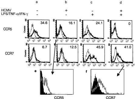
FIG. 3.
CCR5 and CCR7 expression following HCMV infection of DCs. DCs were either mock infected (a and c) or infected with HCMV for 24 h (b and d). DCs then were either left untreated (a and b) (immature DCs) or treated with LPS/TNF-α/IFN-γ for a further 24 h (c and d) (mature DCs). Surface expression of CCR5 or CCR7 was tested by flow cytometry. The broken lines represent the binding of an irrelevant antibody, while the continuous lines represent the binding of CCR5 or CCR7 antibodies, respectively. The numbers represent the percentage of chemokine receptor-expressing cells above background (irrelevant antibody). The results are representative of three experiments. (e and f) CCR5 and CCR7 expression in HCMV-infected cultures on HCMV Ag-positive DCs (black histograms), compared to Ag-negative DCs (gray lines). Two-color flow cytometric analysis of cell surface expression of chemokine receptors and intracellular detection of HCMV pp52 Ag are shown. (e) Expression of CCR5 following HCMV infection of immature DCs. (f) Upregulation of CCR7 by LPS/TNF-α/IFN-γ on TB40/E-infected DCs.

