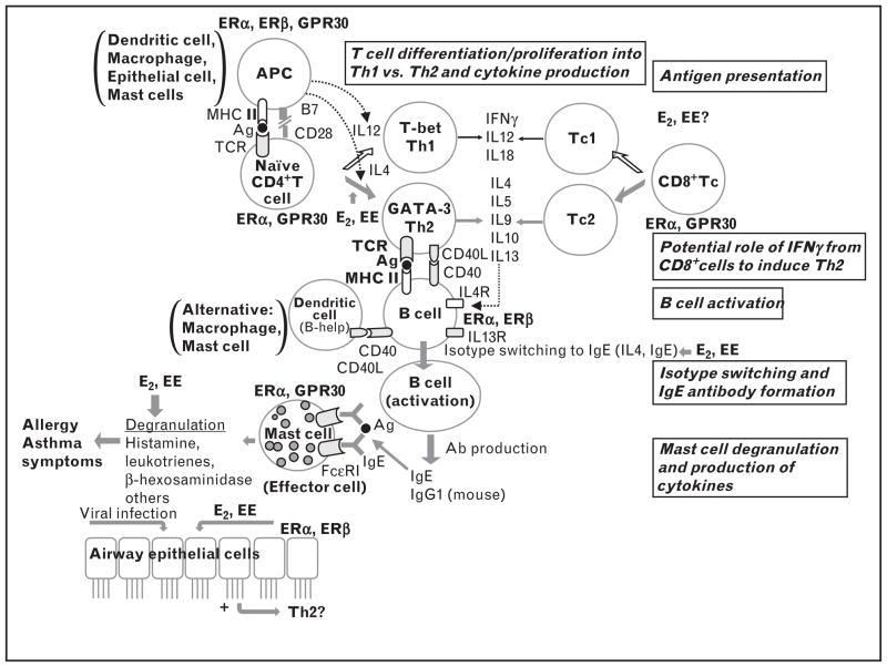
FIGURE 1.
Flow diagram of immune development leading to allergic sensitization with known expression of estrogen receptors by each immune cell type. Environmental estrogens (EEs) have potential effects on each step of allergic sensitization: antigen presentation, Th2 polarization, isotype switching to IgE and mast cell degranulation via ERα, ERβ and G-protein coupled receptor 30 (GPR30) [14,15▪,16–18,19▪▪,20–22,23▪,24,25]. ER, estrogen receptor; IgE, immunoglobulin E.

