Figure 1.

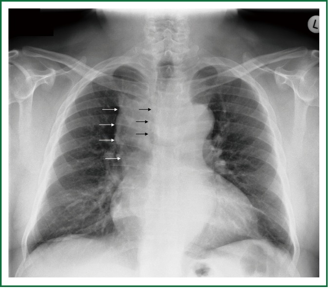
Preoperative chest x-ray. Enlargement of the upper and middle mediastinum (white arrows), mild tracheal deviation to the right, and tracheal stenosis, at the level of the aortic arch (black arrows).

Preoperative chest x-ray. Enlargement of the upper and middle mediastinum (white arrows), mild tracheal deviation to the right, and tracheal stenosis, at the level of the aortic arch (black arrows).