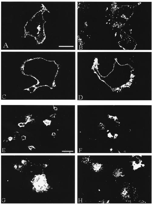
FIG. 1.
Confocal microscopy analysis of gH endocytosis and fusion. HeLa cells were infected with VV-T7 then transfected with gH-wt+gL (A and E), gH-bt+gL (B and F), gH-Y835A+gL (C and G), or gH-S830stop+gL (D and H) and then processed for the confocal endocytosis assay for a 60-min time point (A to D) or the standard confocal assay (E to H). VZV gH was immunolabeled with MAb 258 and goat anti-mouse Alexa 488. Fusion samples (E to H) were examined at a low magnification (×20) to generate landscape images. The bar in panel A is 25 μm and applies to panels A to D; the bar in panel E is 50 μm and applies to panels E to H.

