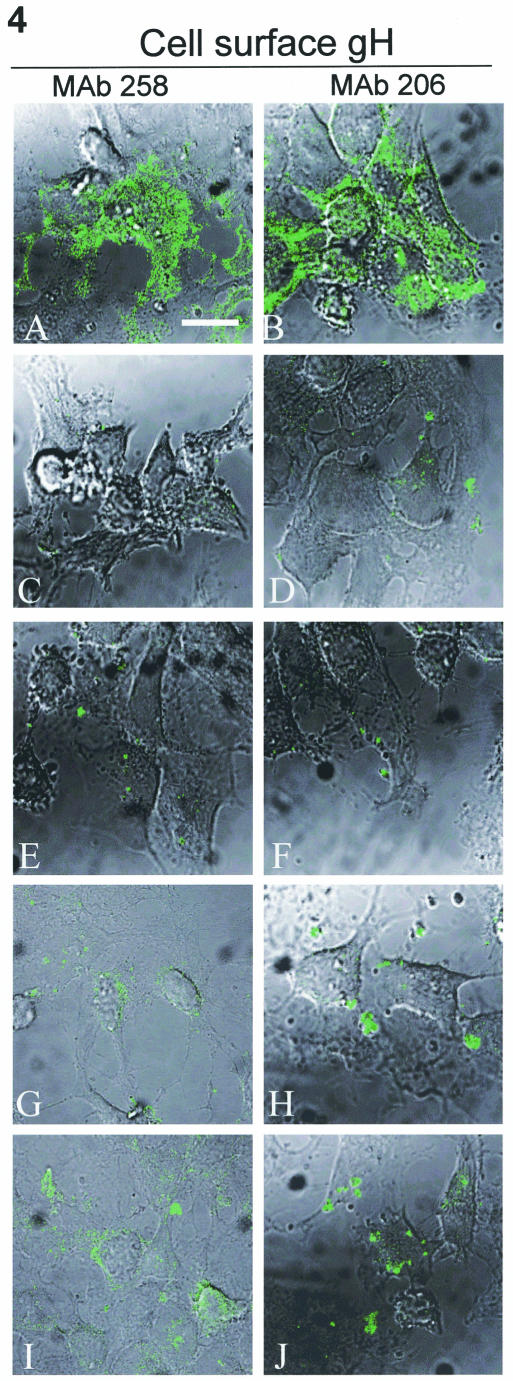
FIG. 4.
Surface expression of immature and mature gH when expressed alone or with gE. HeLa cells were infected with VV-T7 then transfected with gH-wt + gL (A and B), gH-wt (C and D), gH-Y835A (E and F), gH-wt + gE (G and H), or gH-Y835A + gE (I and J) and processed for the standard confocal assay. Samples were fixed without permeabilization to label only surface glycoproteins. Samples were incubated with MAb 258 (A, C, E, G, and I) or MAb 206 (B, D, F, H, and J), followed by goat-anti-mouse Alexa 488. Images were collected with both the fluorescent channel and by Nomarski imaging to show cell morphology. The bar in panel A is 25 μm.

