Figure 2.

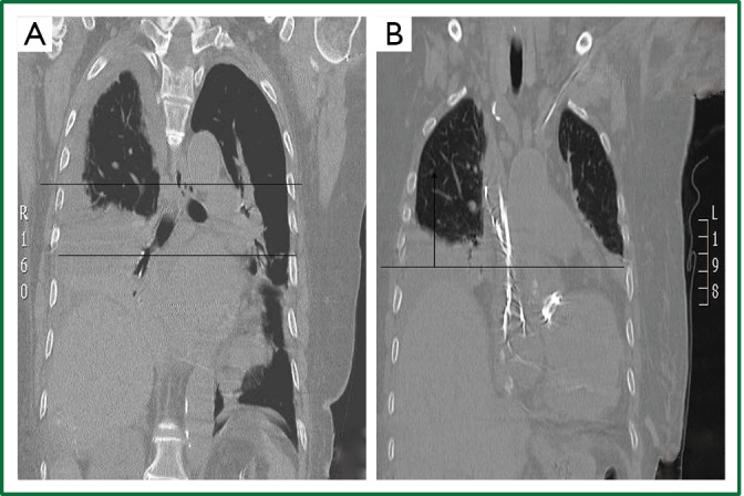
Thoracic CT (coronal plane, supine position). A,B. Elevation of the right henidiaphragm (8 cm above the the left, 2-2.5 intercostal spaces). Lower and middle lobe atelectasis. Pleural effusion (A).

Thoracic CT (coronal plane, supine position). A,B. Elevation of the right henidiaphragm (8 cm above the the left, 2-2.5 intercostal spaces). Lower and middle lobe atelectasis. Pleural effusion (A).