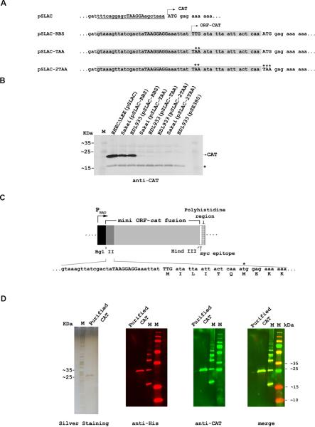
Figure 2. Production of the ORF-CAT fusion protein.
(A) Plasmid sequences. SD2 and the first six codons of the mini-ORF (highlighted) were fused in-frame to the cat gene of pSLAC so as to replace the original SD element of cat. Asterisks mark the mutations subsequently introduced into the mini-ORF-cat gene fusion.
(B) Western blot analysis of ORF-CAT protein synthesis in EHEC transformed with the indicated plasmids. The EHEC strains ΔLEE(pSLAC) and EDL933(pSE380) were used as positive and negative controls, respectively. A non-specific E. coli protein (*) that adventitiously crossreacted with the antibodies was used as a loading control.
(C) Relevant sequence of the 5' region of the mini-ORF-CAT fusion in plasmid pBAD-CAT. The start codon and SD2 are shown in capital letters. The original cat start codon is labeled with an asterisk.
(D) The mini ORF-CAT fusion protein was purified from EHEC cell extracts by metal affinity chromatography on Ni2+-NTA, and its N-terminal sequence was determined (shown in 2C). The purified protein was subjected to SDS-PAGE (left panel) and detected by Western blotting with antisera, as indicated.

