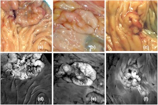
Fig. 7.
Low contrast lesion examples from resected specimens of three patients. (a) Tubulovillous adenoma, (b) Serrated adenoma, (c) Adenocarcinoma. Grayscale ratio images presented in (d)–(f) highlight the lesions which are nonevident in color photos (a)–(c). Dark regions in the lower right corner of image b and center right of image c are caused by tattoo ink applied prior to specimen excision for marking purpose.

