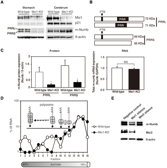
Figure 3. Expression of p21 and m-Numb in the stomach of wild-type and Msi1-KO mice.
(A) Western blots indicating expression of Msi1, p21, and m-Numb protein in the stomach and cerebrum of sham-treated wild-type and Msi1-KO mice. (B) Classification of m-Numb protein by proline-rich region (PRR). (C) m-Numb protein and total RNA expression. Western blotting and quantitative RT-PCR for the expression analysis of m-Numb protein and mRNA in sham-treated wild-type and Msi1-KO mice was performed in triplicate. The density of each m-Numb protein band in western blotting is normalized to actin and represented as the fold-change relative to expression of the protein in wild-type mice. White bars: wild-type mice; black bars: Msi1-KO mice. *P<0.01 compared to wild-type mice. (D) Polysome analysis of m-Numb mRNA from mouse stomach. Fractions of a polysome gradient prepared from the stomachs of wild-type (empty circles) and Msi1-KO (filled circles) mice. RNA was extracted from each fraction and used for quantitative RT-PCR. The results are shown as the percentage of the total amount of RNA in each fraction. (E) Knockdown of human Msi1 and Msi2 in N87 cells by shRNA-containing lentiviral particles. Western blotting was performed using primary antibodies specific for Msi1, Msi2, m-Numb, and β-actin.

