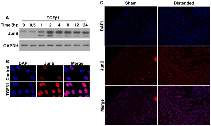
Figure 2. JunB levels are increased in BSMC in response to TGFβ1, and in an ex vivo model of rodent bladder distension.
(A) BSMC were treated with TGFβ1 for the indicated times and assessed for JunB levels by immunoblotting. GAPDH is included as a loading control. (B) Immunofluorescence analysis of BSMC showing increased JunB nuclear localization upon TGFβ1 treatment for 24 h. (C) Sections from rat bladders distended ex vivo for 8 h (injured) were stained sequentially with anti-JunB and Cy3-conjugated species-specific secondary antibody. Increased nuclear fluorescent signal for both proteins was evident in the detrusor smooth muscle of stretch-injured specimens, but not of non-distended (control) bladders.

