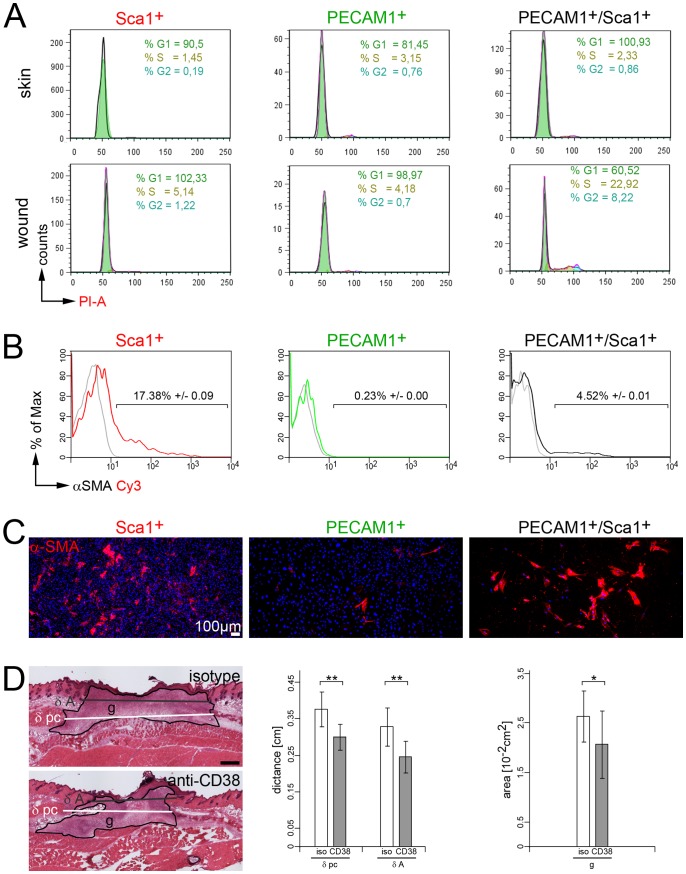
Figure 5. Myofibroblast-like cell formation and modulation of CD38 receptor activity. (A).
Representative cell cycle analysis of skin- and wound-derived Sca1+, PECAM1+ and PECAM1+/Sca1+ cells seven days post injury using propidium iodide (PI) stain in flow cytometry analysis (see figure S1). The relative percentage of cells in G1 (green), S (ochre) and G2 (blue) are highlighted. (B) Flow cytometric detection of α-SMA in wound-derived Sca1+, PECAM1+/Sca1+ and PECAM1+ cells seven days post injury (n = 7 mice). (C) Immunofluorescence analysis of α-SMA expression in cultured wound-derived Sca1+, PECAM1+ and PECAM1+/Sca1+ cells. Nucleoli were detected using DAPI stain. (D) Morphometric analysis of wounds in immunodeficient mice stimulated with rat anti-CD38 or isotype matched antibodies (n = 4). Distances between edges of the panniculus carnosus (δ pc), hair follicles (δ A) and the area of the granulation tissue (g) were determined. Statistics: unpaired two-tailed student’s T-test (*p≤0.05, **p≤0.01). Bars 100 µm (C), 500 µm (D).

