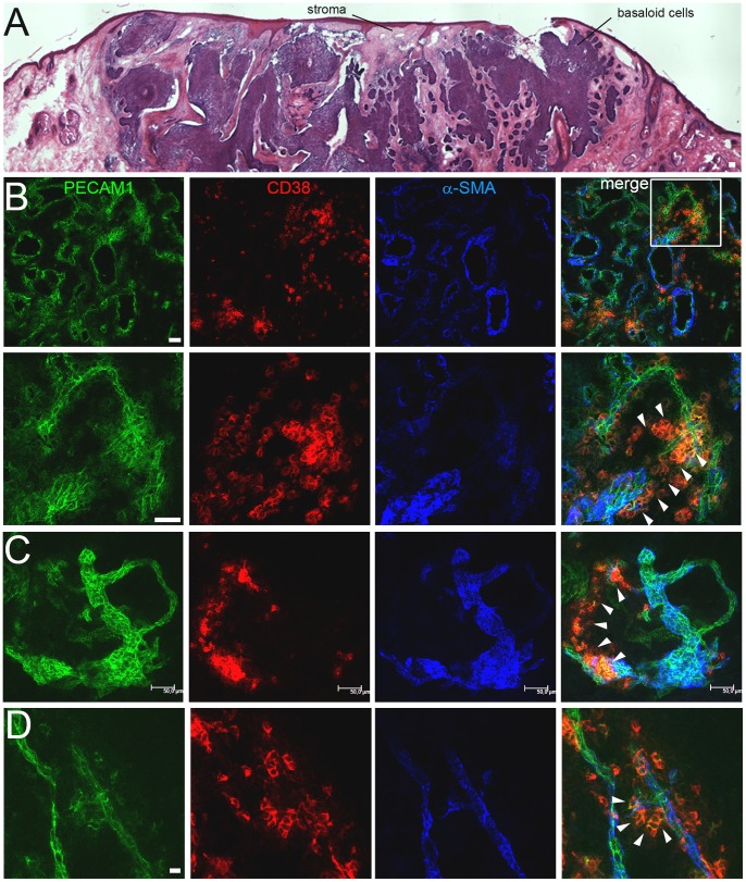
Figure 7. Identification of PECAM1+/CD38+ cells in human basal cell carcinomas (BCC).
(A) H&E-stained cryosection of a BCC. (B–D) Confocal microscopy analysis of PECAM1, CD38 and α-SMA expression in two BCC biopsies (B–C, D). (B) Overview (top row) of the highly vascularized PECAM1+ stroma. Squares within the merged image indicate the magnified region (lower row). PECAM1low/CD38+ cells are marked (arrowheads). Bars 100 µm (A), 50 µm (B, C), 10 µm (D).

