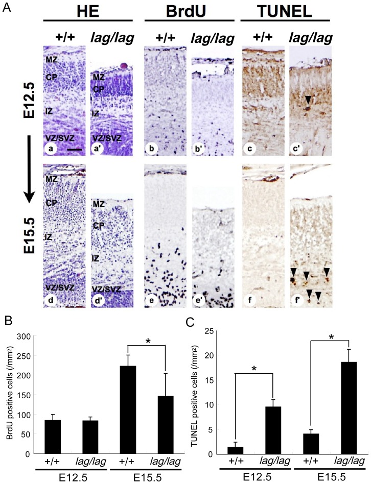
Figure 10. The increased apoptosis and the decreased cell proliferation during development of the cerebral cortex in lag/lag mice.
(A) Pregnant mice were intraperitoneally injected with BrdU 2 h before sacrifice. Littermate wild type (+/+) (Aa, Ab, Ac, Ad, Ae, Af) and mutant (lag/lag) (Aa’, Ab’, Ac’, Ad’, Ae’, Af’) cerebral cortex coronal sections at E12.5 and 15.5 were stained with hematoxylin, anti-BrdU antibody and TUNEL. CP, cortical plate; IZ, intermediate zone; MZ, marginal zone; VZ/SVZ, ventricular zone/subventricular zone. Bars, 100 µm. (B) The number of BrdU immuno-positive cells (n = 4). Error bars represent SD. Asterisk indicates statistical significance (t test; *, p<0.05). (C) The number of TUNEL immuno-positive cells in (n = 4). Cell counts were performed in the sensory-motor cortex. Error bars represent SD. Asterisks indicate statistical significance (t test; *, p<0.01).

