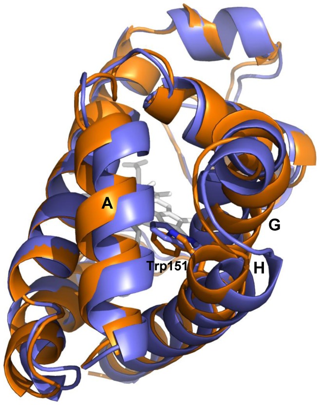
Figure 5. Structural differences in the two species of O2Cygb.
Superposition of the average structures derived for the two oxygenated forms of Cygb (built up using as templates the X-ray structures of HE7Q mutant and 3AG0, which are shown in violet and orange, respectively). The helices that surround Trp151 are also labelled.

