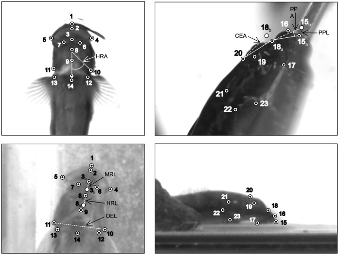
Figure 1. Still images of S. stimpsoni in (a) ventral and (b) lateral views, illustrating anatomical landmarks that were digitized to generate kinematic data.
For ventral view (a), labeled points are as follows: (1) anterior edge of upper lip, (2) anterior tip of inner edge of upper lip, (3) anterior tip of mandibular symphysis, (4) right caudo-lateral tip of mouth, (5) left caudo-lateral tip of mouth, (6) midpoint on right side of mandible between mandibular symphysis and right caudo-lateral tip, (7) midpoint on left side of mandible between mandibular symphysis and left caudo-lateral tip, (8) hyoid arch, (9) midline joint between left and right branchiostegal rays, (10) caudolateral margin of right operculum, (11) caudolateral margin of left operculum, (12) right pectoral fin base, (13) left pectoral fin base, and (14) anterior tip of pelvic sucker. For lateral view (b), labeled points are as follows: (15) anterior tip of upper lip, (16) anterior edge of upper lip base, (17) caudal tip of junction between maxilla and dentary, (18) anterior edge of neurocranium, (19) center of eye, (20) junction between neurocranium and epaxial muscle insertion, (21) caudal edge of operculum, (22) dorsal edge of pectoral fin base, and (23) ventral edge of pectoral fin base spine.

