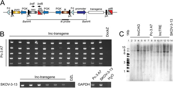
Figure 3.
Fidelity of the docking-incoming system. (A) Position of the primer pairs used for screening of integration (IntF and IntR) as well as the tk probe used for southern analysis. (B) PCR amplification of the integration junction using primers recognizing the attL site and the neoR probe. Sixty-six colonies (60 colonies for Pc-3-A7 and 6 for SKOV-3-13) were screened and all had the correct integration site. Analysis was done using a multicomp agarose gel. (C) Southern blot analysis of subclones derived by integration of two different incoming vectors (IncCAG-transgene; lanes 2–7, and IncTRE-transgene; lanes 9–13) into line Pc-3-A7 (lanes 2–7) and SKOV-3-13 (lanes 9–13). Genomic DNA was digested with BamHI and the tk probe was used. Original Pc-3-A7 and SKOV3-13 DockZ lines were also included in lanes 8 and 14, respectively. 1-Kb marker is shown in the first lane.

