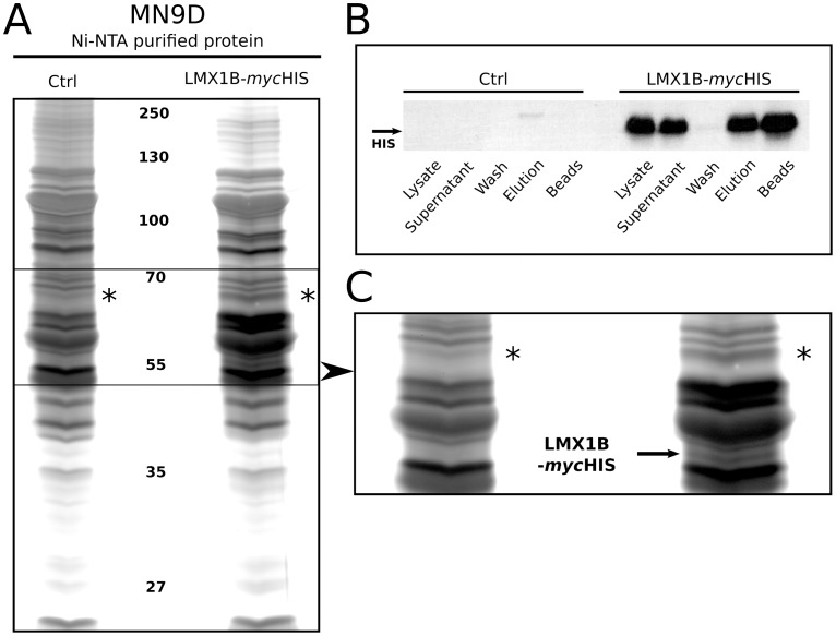
Figure 1. Affinity purified Lmx1b-HIS proteins.
(A) Lmx1b-HIS purified proteins from MN9D cells, by means of HIS tagged affinity purification via Ni-NTA agarose beads, followed by separation on silver-stained SDS gel. The left lane represents proteins purified from control transfected MN9D cells. The right lane shows purified proteins from LMX1B-HIS overexpressing MN9D cells. Asterisks mark an observed differential protein band. (B) Western blot validation of the LMX1B-HIS overexpression in MN9D cells, followed by successful purification of LMX1B-HIS protein. Lysate shows clear LMX1B-HIS overexpression. Supernatant reveals unbound LMX1B-HIS after Ni-NTA bead incubation. Protein is strongly bound to the beads, as shown by an extremely low amount of protein that was detected in the first washing-steps. Following successful elution, beads were heated and used for a second, thorough elution; a large amount of protein was detected that was not eluted in the first elution step. (C) Detailed image of the differential protein band in the LMX1B-HIS OE sample (asterisks). Overexpressed LMX1B-HIS protein (based on size) was detected as well (arrow). Ctrl, purified protein from control transfected MN9D cells; LMX1B-mycHIS, purified protein from MN9D cells overexpressing LMX1B-HIS.

