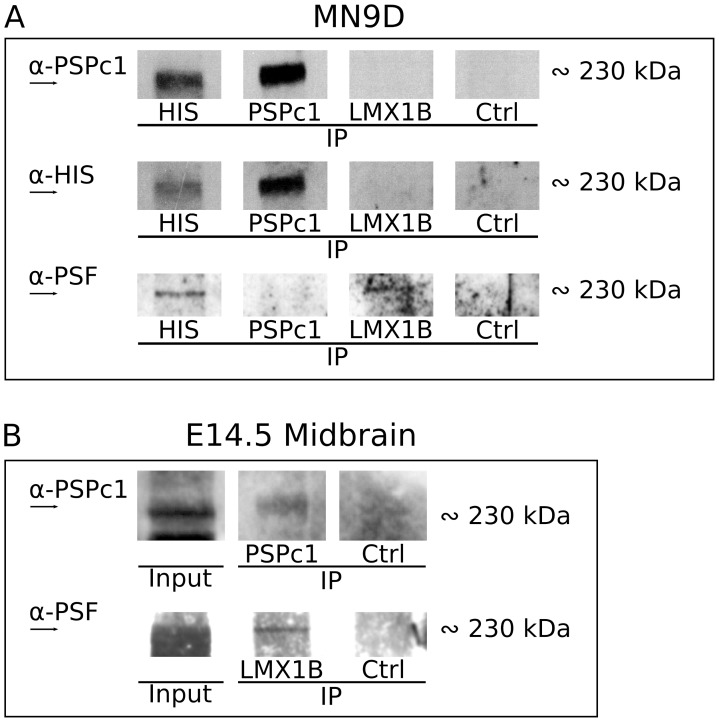
Figure 5. Identification of a 230 kDa complex containing LMX1B(-HIS), PSPC1 and PSF.
(A) PSPC1 protein is identified in a protein band of approximately 230 kDa, in a PSPC1 IP and in a HIS IP, but not in an LMX1B IP. When immunoblotting for HIS, a protein of the same size is detected in these IP experiments, and again not in an LMX1B IP. After immunoblotting for PSF, of the same blot, PSF is detected at 230 kDa in the HIS IP, faintly in the PSPC1 IP, and in the LMX1B IP. (B) Analysis of 230 kDa proteins in vivo, in E14.5 dissected midbrain tissue. PSPC1 pull-down reveals PSPC1 protein at 230 kDa, suggesting that the 230 kDa protein complex exist in vivo. At the same height, PSF is detected when immunoprecipitating for LMX1B. Ctrl, control IP with normal host serum; IP, immunoprecipitation.

