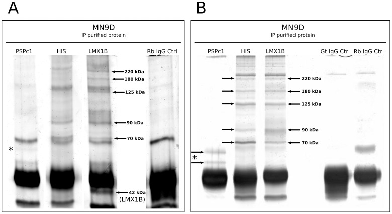
Figure 6. LMX1B co-immunoprecipitated protein identification.
(A) IP against HIS, PSPC1 and LMX1B on LMX1B-HIS OE MN9D cell lysate, followed by separation on silver-stained SDS gel. The PSPC1 IP shows two protein bands that might represent both PSPC1 isoforms. Five protein bands of the LMX1B IP were excised and used for mass spectrometry analysis (arrows). In addition, the 42 kDa protein was taken along, as a positive control, and was later confirmed as LMX1B protein. (B) Second IP experiment against HIS, PSPC1 and LMX1B on LMX1B-HIS OE MN9D cell lysate, followed by separation on silver-stained (FireSilver kit, Proteome Factory, Berlin) SDS gel, confirming and improving the pattern as described in the first IP experiment (A). The PSPC1 IP shows two protein bands that likely represent both PSPC1 isoforms, and they were used for mass spectrometry analysis. Five differential protein bands of the LMX1B and HIS IP were excised and used mass spectrometry analysis (arrows). Gt IgG Ctrl, goat IgG control; Rb IgG Ctrl, rabbit IgG control.

