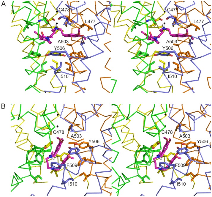
Figure 5. Detailed stereo view of the ivabradine docking in the hHCN4 WT channel.
Stereo view of the interior of the WT hHCN4 channel in its closed (A) and open (B) form with the best-docked pose of ivabradine shown in magenta. The S5-P-S6 regions of the four hHCN4 subunits are shown as ribbon in green yellow, orange and pale blue, respectively. Side-chains of residues interacting with the ivabradine molecule are shown in stick representation in both panels. For the closed state only, the main-chain atoms of C478 are shown since the carbonyl oxygen atoms may form additional H-bonds with ivabradine. I510 is also shown in the channel closed form, though this residue does not interact directly with ivabradine. Black spheres indicate positions corresponding to K+ ion bound in pore of the KcsA crystal structure [12]. For clarity only residues of one subunit are labeled and L477 of the pale blue subunit is omitted.

