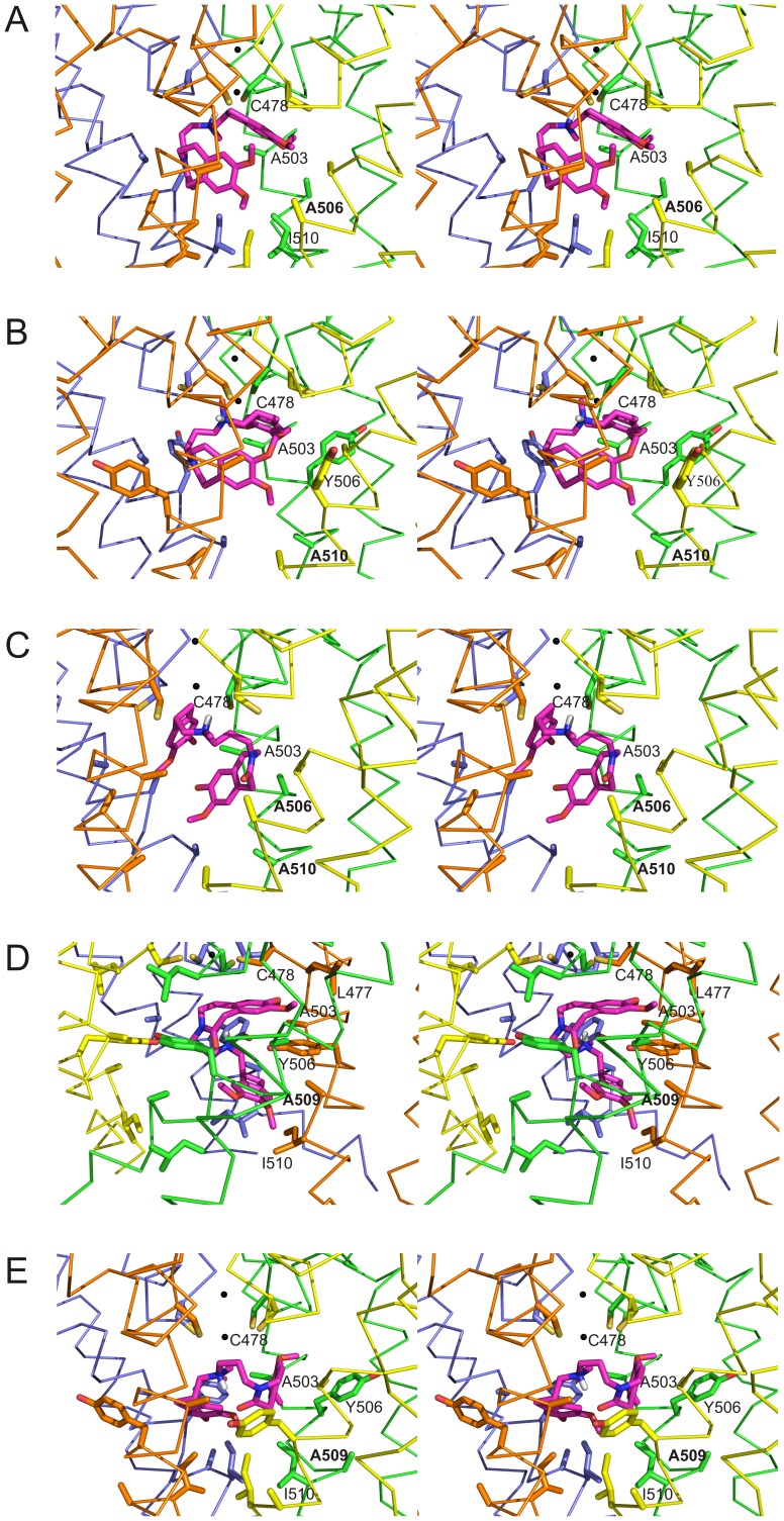
Figure 6. Detailed stereo view of the ivabradine docking to hHCN4 mutant channels.
A,B,C: stereo views of the interior of hHCN4 mutants Y506A (A), I510A (B), and Y506A-I510A (C) in the closed form. D, E: stereo views of the interior of the F509A mutant in the open (D) and closed (E) form. Side-chains of residues relevant to ivabradine binding are shown as ball-and-stick in all panels. In all mutant models the best pose of the docked ivabradine is shown in magenta, while the S5-P-S6 regions of the four hHCN4 subunits are shown as ribbon in grey, yellow, orange and pale blue, respectively. For clarity, residues have been labelled only in one hHCN4 subunit, with mutated residues indicated in bold characters.

