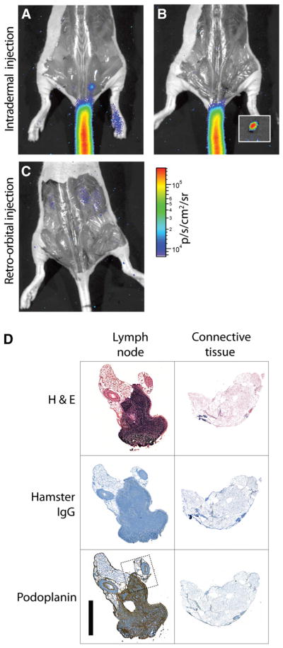
Figure 6.
Cerenkov-guided surgical resection of 18F-FDG–bearing LN, 10 min after injection, with surgical validation. (A) Lateral-tail intradermal injection yields greater uptake in 1 sacral node, seen with just skin removed. PET image is included as Supplemental Figure 2. (B) CR guides resection of node, magnified in inset. (C) Systemic administration of 18F-FDG (retroorbital injection) does not enable identification of nodes, even after surgical exposure. Signal from renal clearance can be seen. (D) Immunohistologic verification of Cerenkov-guided excised tissues. Resected node as indicated by CR, along with surrounding connective tissue, was stained with hematoxylin and eosin, control hamster IgG, and podoplanin. The stromal region of node is indicated by dense hematoxylin and eosin and positive podoplanin staining. Dotted line indicates region of magnification (included as Supplemental Fig. 4). Scale bar, 1 mm. H&E = hematoxylin and eosin.

