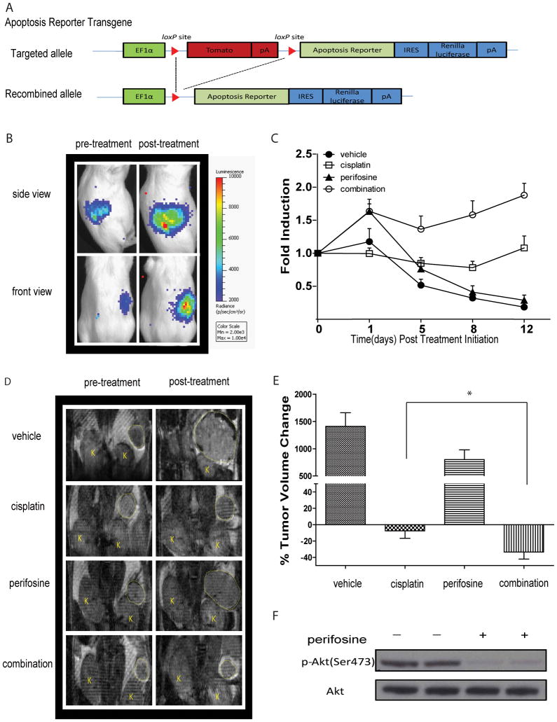
Figure 4. Generation of new reporter OEA model identifies AKT as molecular target for therapy.
(A) Schematic of the bioluminescence apoptosis reporter transgene. EF-1 alpha drives expression of floxed fluorescence protein tdTomato, which is removed upon tissue specific Cre recombination. Conditional Cre recombination leads to transcription of the bioluminescent apoptosis reporter and IRES dependent renilla luciferase expression. (B) Representative bioluminescence images from Apc−/−, Pten −/−, Apopreporter+ animals before treatment and one day after treatment. Bioluminescence signal is detected in the right but not left ovaries of animals. (C) When tumor size reached ≥ 50 mm3 animals were randomized into four treatment groups and BL- and MR- imaging was performed before treatment and on day1, day 5, day 8 and day 12 post treatment initiation. Fold induction was calculated by normalizing bioluminescence signals to tumor volumes acquired by MRI and pre-treatment values at each time point. Data represents mean ± SEM. (D) Representative MR images of animals in each group pre- and two weeks post treatment. (E) Percent change in tumor volume as measured by MR imaging. Data represents mean ± SEM. Statistical significance was assessed at a p<0.05 (*) using an unpaired Student’s t-test. (F) Representative images of western blotting analysis for total AKT and pAKT (Ser473) 3 hours post treatment with perifosine of tumors obtained from Apc−/−, Pten −/−, Apopreporter+ animals.

