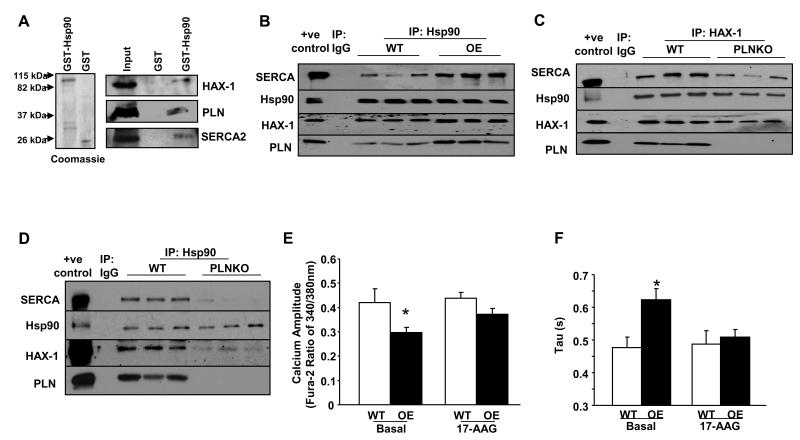
Figure 8. Hsp90 associates with PLN/SERCA.
A: SDS-gel stained with Coomassie blue showing purified GST and GST-Hsp90 recombinant proteins. GST-pull down assays were performed using mouse cardiac homogenates and GST-Hsp90 or GST recombinant proteins. The association with SERCA2, HAX-1 and PLN was determined by western blot analysis with appropriate antibodies. B: Centrifuged homogenates from WT and HAX-1 OE hearts were subjected to co-immunoprecipitation using the Hsp90 antibody. Five independent experiments were performed. C and D: Same procedure (as B above) was performed with WT and PLNKO homogenates, using HAX-1 and Hsp90 antibodies. Five independent experiments were performed. E and F: Calcium transient peak (n= 47-73 cells from 9 WT hearts and 8 HAX-1 OE hearts) and time constant for calcium decay (Tau, n=16-30 cells from 4 hearts in each group) in isolated WT and HAX-1 OE myocytes with or without the administration of 17-AAG. *P<0.05, compared to WT basal. Data are presented as mean±SEM.

