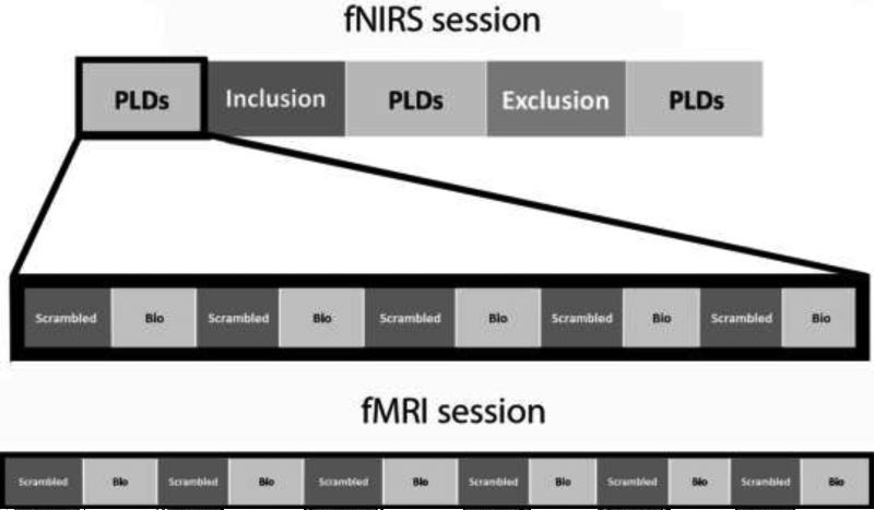
Figure 1.
Visual depiction of the experimental paradigm for the fNIRS session (top) and fMRI session (bottom). Each block of biological (bio) and scrambled motion consisted of a 24-second point-light video clip. The fNIRS session consisted of three runs of 10 PLD videos, presented at baseline, post-inclusion, and post-exclusion. The fMRI session consisted of one run of 12 PLD videos.

