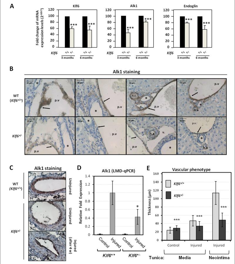
Figure 3. Klf6+/− heterozygous mice express lower levels of Alk1 in both basal condition and after endothelial injury.
A. Real time RT-PCR of Alk1, Klf6 and endoglin levels from total liver mRNA of Klf6+/− heterozygous mice (3 and 6 months-old) compared to their wild type siblings. B. Immunohistochemistry of Alk1 in hepatic vasculature of Klf6+/− and Klf6+/+ mice livers. Arrows highlight the Alk1 staining in ECs. The asterisks indicate the bile ducts. h.a, hepatic artery; p.v, portal vein; v, vein. C. Immunohistochemical staining of Alk1 protein in 4 weeks-injured femoral arteries from Klf6+/− mice in comparison with wild type littermates. D. Quantification of Ak1 mRNA by qPCR using laser microscopy microdissection (LMD) from tissue sections of femoral arteries. E. Measurement of the tunica media and neointima 4 weeks post-injury. Each value represents the mean of at least 75 different measurements.

