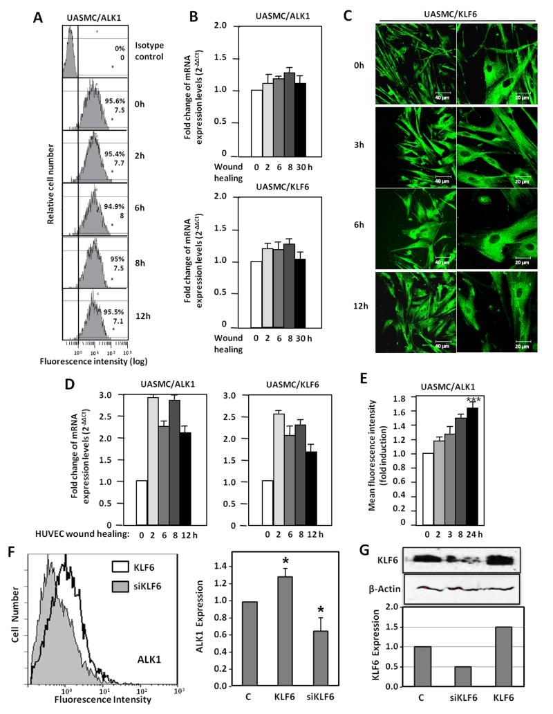
Figure 8. Paracrine effect of HUVEC denudation on ALK1 expression in vascular SMCs.
A. ALK1 expression in the surface of vSMCs after denudation in vitro, measured by flow cytometry. B. Real time RT-PCR of ALK1 and KLF6 after vSMC denudation. C. Immunofluorescent staining of KLF6 in vSMCs during wound healing. D,E. Effect of conditioned media from HUVEC subjected to denudation at different time points on vSMC treated overnight. D. Real time RT-PCR of ALK1 and KLF6 transcripts. E. ALK1 expression measured by flow cytometry and represented as fold induction. F,G. Effect of conditioned media from HUVEC subjected to KLF6 overexpression (pCiNeoKLF6; KLF6), KLF6 suppression (pSuper-siKLF6; siKLF6) or mock transfection (C) on vSMC treated overnight. F. ALK1 expression measured by flow cytometry (left) and represented as fold induction (right). G. Western blot analysis of KLF6 in total cell lysates. The intensity of the KLF6 band relative to β-actin intensity is represented in the histogram. UASMC, umbilical artery smooth muscle cells.

