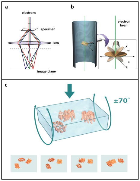
Figure 1.
Image formation in the electron microscope. (a) Schematic illustrating image formation in an electron microscope, highlighting the similarities between electron and optical microscopy. (b) Schematic illustrating the principle of data collection for electron tomography. As the specimen is tilted relative to the electron beam, a series of images is taken of the same field of view. (c) Rendering of selected projection views generated during cryo-electron tomography as a vitrified film (formed by rapidly freezing a thin aqueous suspension) is tilted relative to the electron beam. To reconstruct the three-dimensional volume, a set of projection images is “smeared” out along the viewing directions to form back-projection profiles. The images are combined computationally to recover the density distribution of the object. Figure adapted from [110].

