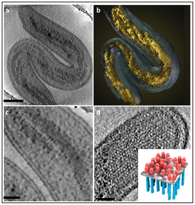
Figure 2.
Use of cryo-electron tomography to image the interior architecture of intact bacterial cells. (a,b) Illustration of spiral architecture of the nucleoid in Bdellovibrio bacteriovorus showing (a) a 210 Å thick tomographic slice through the 3D volume of a cell and (b) a 3D surface rendering of the same cell, with the spiral nucleoid highlighted (yellow). (c) Higher magnification view of a tomographic slice through the cell, showing well-separated nucleoid spirals and ribosomes (dark dots) distributed at the edge of the nucleoid. (d) Expanded views of 210 Å thick tomographic slices, showing top-views of polar chemoreceptor arrays. A schematic model (inset) illustrates the spatial arrangement of the chemoreceptor arrays in the plane of the membrane. Scale bars: 2000 Å in (a) and 500 Å in (c,d). Figure adapted from [44].

