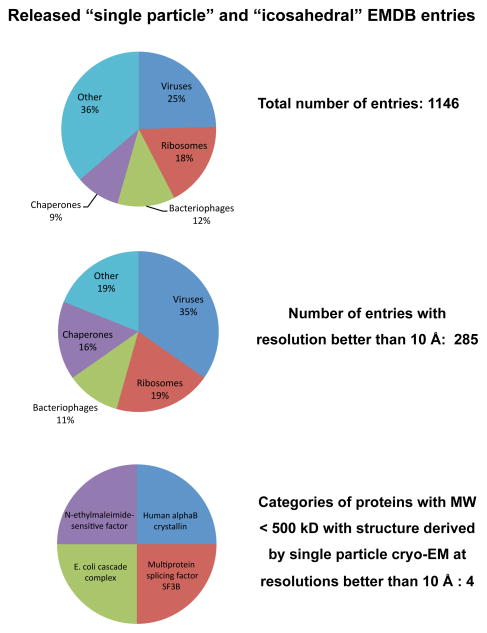
Figure 9.
Distribution of released entries belonging to the “single-particle” and “icosahedral” categories in the Electron Microscopy Data Bank (emdb.org) as of June 2012. Structures from ribosomes and icosahedral viruses dominate the deposited entries, both in the entire data set, and even more strikingly, in the subset of entries with resolutions better than 10 Å. Further inspection shows that there are only four distinct protein complexes with molecular masses < 500 kDa (Multiprotein splicing factor SF3B: EMD-1043; DegQ: EMD-5290; E. coli cascade complex: EMD-5314 and EMD-5315; N-ethylmaleimide-sensitive factor: EMD-5370 and EMD-5371), thus identifying a key gap in structural biology that can potentially be filled by taking advantage of improvements in microscope hardware and algorithms for image processing.

